Understanding the Generalized Animal Cell: Generalized Animal Cell Coloring Sheet
Generalized animal cell coloring sheet – Animal cells are the fundamental building blocks of animal tissues and organs. They are eukaryotic cells, meaning they possess a membrane-bound nucleus and other organelles, unlike their prokaryotic counterparts. Understanding their structure and function is crucial to comprehending the complexities of animal life.
Generalized animal cell coloring sheets offer a great way to learn about biology, providing a visual representation of cellular structures. For a different kind of creative outlet, consider the vibrant illustrations in the disney junior animated coloring book , which offers a fun contrast to the scientific accuracy of the cell diagrams. Returning to the animal cell, remember that accurate coloring helps solidify understanding of organelles and their functions.
Basic Components and Their Functions
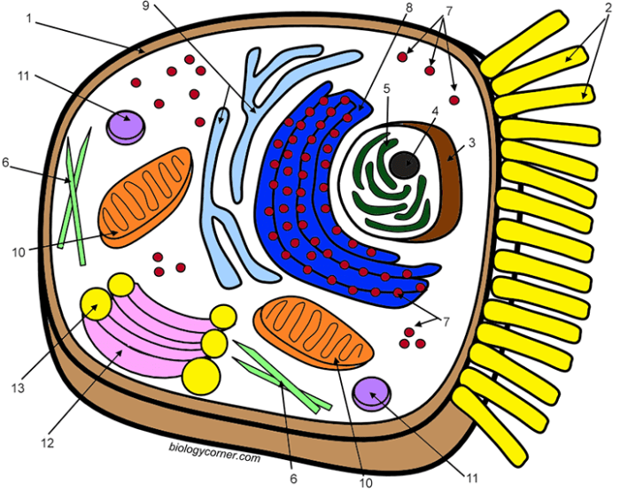
The generalized animal cell comprises several key components, each with a specific role in maintaining cellular life. The nucleus, for example, houses the cell’s genetic material (DNA), controlling all cellular activities. The cytoplasm, a gel-like substance filling the cell, contains various organelles. Mitochondria, often referred to as the “powerhouses” of the cell, generate energy through cellular respiration.
Ribosomes are responsible for protein synthesis, translating genetic information into functional proteins. The endoplasmic reticulum (ER), a network of membranes, plays a vital role in protein and lipid synthesis and transport. The Golgi apparatus modifies, sorts, and packages proteins for secretion or delivery to other parts of the cell. Lysosomes contain enzymes that break down waste materials and cellular debris.
Finally, the cell membrane, a selectively permeable barrier, regulates the passage of substances into and out of the cell.
Differences Between Plant and Animal Cells
While both plant and animal cells are eukaryotic, several key differences distinguish them. Plant cells possess a rigid cell wall made of cellulose, providing structural support and protection, which is absent in animal cells. Plant cells also contain chloroplasts, the organelles responsible for photosynthesis, enabling them to produce their own food. This capability is lacking in animal cells, which rely on consuming other organisms for energy.
Finally, plant cells typically have a large central vacuole for storage of water and other substances, while animal cells may have smaller vacuoles or none at all.
Cellular Processes: Respiration and Protein Synthesis
Cellular respiration is the process by which cells convert glucose and oxygen into ATP (adenosine triphosphate), the cell’s primary energy currency. This process occurs in the mitochondria, involving a series of complex chemical reactions. Protein synthesis, on the other hand, is the process of creating proteins from the information encoded in DNA. This process involves transcription (copying the DNA sequence into mRNA) in the nucleus and translation (assembling amino acids into a polypeptide chain according to the mRNA sequence) on ribosomes, often with the assistance of the endoplasmic reticulum and Golgi apparatus.
Labeled Diagram of a Generalized Animal Cell
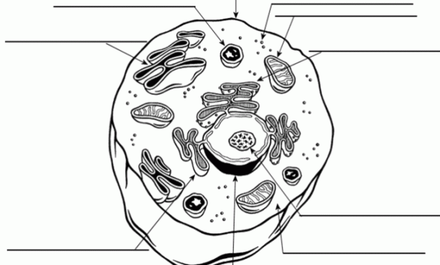
Imagine a circle representing the cell membrane. Within this circle, a slightly smaller, irregular circle represents the nucleus, containing the nucleolus (a smaller, denser region within the nucleus). Scattered throughout the cytoplasm (the space between the cell membrane and the nucleus) are numerous smaller ovals representing mitochondria. A network of interconnected, branching lines represents the endoplasmic reticulum, with some regions appearing rough (due to ribosomes attached) and others smooth.
Near the nucleus, a stack of flattened sacs represents the Golgi apparatus. Small, oval structures scattered throughout the cytoplasm represent lysosomes. Finally, tiny dots scattered throughout the cytoplasm and on the rough endoplasmic reticulum represent ribosomes. The entire structure is enclosed by the cell membrane.
Designing the Coloring Sheet Layout
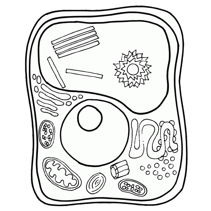
Creating a visually appealing and informative animal cell coloring sheet requires careful consideration of layout and design elements. The goal is to present a clear and engaging representation of the cell’s structure, facilitating understanding and enjoyment for the user. The layout should be both aesthetically pleasing and functionally effective in conveying complex biological information in a simplified manner.The optimal dimensions for a printable coloring sheet would be 8.5 x 11 inches (standard letter size), allowing for ample space without excessive blank areas.
This size is widely compatible with home and school printers. Larger dimensions could be considered for more detailed depictions, but would require more paper and potentially increased printing costs.
Layout Options for Varying Detail Levels, Generalized animal cell coloring sheet
Different layout options can cater to various age groups and learning levels. A simplified version might focus on the major organelles – the nucleus, cytoplasm, cell membrane, and perhaps a few others – using large, easily colorable shapes. This approach is suitable for younger children. More complex versions could incorporate additional organelles such as mitochondria, ribosomes, Golgi apparatus, endoplasmic reticulum, and lysosomes, using smaller, more detailed shapes and potentially including labels for each organelle.
This more detailed version would be appropriate for older children or students studying cell biology. A key consideration is the balance between visual clarity and the inclusion of relevant detail.
Organelle Placement and Logical Organization
The arrangement of organelles within the cell should be logical and intuitive. The nucleus, typically the largest organelle, should be centrally located. Other organelles can be positioned around the nucleus, reflecting their relative size and spatial relationships within a real cell. For instance, the endoplasmic reticulum could be depicted as a network extending from the nucleus, while mitochondria could be scattered throughout the cytoplasm.
Using visual cues like arrows or connecting lines to show relationships between organelles could enhance understanding. The cell membrane should clearly define the outer boundary of the cell.
Whitespace and Visual Appeal
Effective use of whitespace is crucial for improving readability and visual appeal. Sufficient spacing around organelles prevents overcrowding and improves the clarity of individual components. Whitespace also allows for the addition of labels or text without cluttering the drawing. The overall layout should be balanced, avoiding a cramped or uneven distribution of elements. Consider using a light background color or a subtle border to further enhance the visual appeal and make the coloring sheet more engaging.
Presentation and Formatting
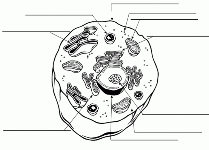
Effective presentation is crucial for a successful educational coloring sheet. A well-structured layout enhances understanding and engagement, making the learning process more enjoyable and memorable. This section details formatting choices for optimal impact.
Organelle Comparison Table
A comparative table provides a concise overview of various cell organelles. This visual aid allows for quick comprehension of their functions and locations. The table below uses four responsive columns to ensure readability across various screen sizes.
| Organelle | Function | Location within Cell | Description |
|---|---|---|---|
| Nucleus | Contains the cell’s genetic material (DNA) and controls cell activities. | Center of the cell | Think of it as the cell’s control center, directing all operations. The DNA inside dictates what the cell does and how it grows. |
| Mitochondria | Generate energy (ATP) through cellular respiration. | Throughout the cytoplasm | These are the cell’s powerhouses, converting nutrients into usable energy. They are often depicted as bean-shaped. |
| Ribosomes | Synthesize proteins. | Free-floating in cytoplasm or attached to the endoplasmic reticulum | These tiny structures are the protein factories of the cell. They translate genetic instructions into functional proteins. |
| Endoplasmic Reticulum (ER) | Synthesizes lipids, modifies proteins, and transports materials. Rough ER has ribosomes attached; smooth ER does not. | Network extending throughout the cytoplasm | The ER is a complex network of membranes involved in a variety of cellular processes. The rough ER is involved in protein synthesis, while the smooth ER plays a role in lipid metabolism. |
| Golgi Apparatus | Processes, packages, and distributes proteins and lipids. | Near the nucleus | Think of this as the cell’s post office; it modifies and packages proteins for transport to their final destinations. |
| Lysosomes | Break down waste materials and cellular debris. | Throughout the cytoplasm | These are the cell’s recycling centers, responsible for breaking down unwanted materials. They contain digestive enzymes. |
| Cell Membrane | Regulates the passage of substances into and out of the cell. | Outer boundary of the cell | The cell membrane acts as a selectively permeable barrier, controlling what enters and exits the cell. |
Incorporating the Coloring Sheet into Educational Resources
The coloring sheet can be integrated into a broader lesson plan by using it as a pre-activity to introduce cell organelles, a post-activity to reinforce learning, or as a supplementary activity for visual learners. It could also be incorporated into a science fair project, a classroom activity to be completed individually or in groups, or as part of a homework assignment.
Following the coloring activity, students can label the organelles, write short descriptions of their functions, or even create a short presentation summarizing their knowledge.
Presenting the Completed Coloring Sheet in a Science Project
The completed coloring sheet, with neatly labeled organelles, can serve as a visual aid in a larger science project. It could be included as part of a presentation, a poster, or a report. The project could focus on the overall structure and function of the animal cell, comparing and contrasting animal cells with plant cells, or exploring specific organelles in greater detail.
The coloring sheet provides a concrete visual foundation for more advanced learning.
