Introduction to Plant and Animal Cell Coloring Activities
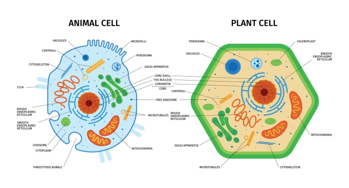
Plant and animal cell coloring – Coloring plant and animal cells isn’t just a fun activity; it’s a powerful educational tool! By engaging with the visual representation of these tiny building blocks of life, students can develop a deeper understanding of their structures and functions. The act of coloring helps solidify knowledge and makes learning more memorable and enjoyable.The visual differences between plant and animal cells are significant and readily apparent through coloring.
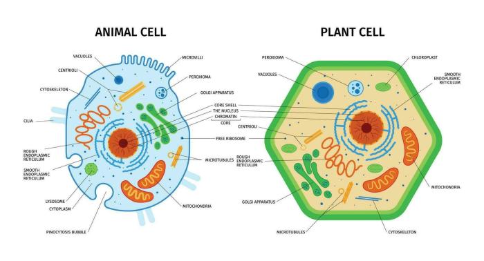
Plant cells possess a rigid cell wall, a large central vacuole, and chloroplasts, features absent in animal cells. Animal cells, on the other hand, are typically more irregularly shaped and lack these defining structures. Coloring these differences highlights the unique characteristics of each cell type and helps students visually distinguish between them.
Materials Needed for Plant and Animal Cell Coloring
Gathering the necessary materials ensures a smooth and engaging coloring experience. Having everything prepared beforehand minimizes interruptions and allows for focused learning. The following table lists essential items:
| Item | Description | Quantity | Notes |
|---|---|---|---|
| Printable Worksheets | Worksheets showing Artikels of plant and animal cells, labeled with key organelles. | 1-2 per student | Easily found online or created using cell diagrams. |
| Colored Pencils | A variety of colors for differentiating cell components. | 1 box per student | Crayons or markers can also be used. |
| Ruler | For precise labeling and drawing. | Optional, but helpful | Helps with neatness and accuracy. |
| Sharpener | To keep colored pencils pointed for detail. | Optional, but recommended | Ensures clear coloring and labeling. |
Key Differences in Plant and Animal Cell Structures
Plant and animal cells, while both eukaryotic, meaning they have a membrane-bound nucleus and other organelles, exhibit significant structural differences that reflect their distinct functions and lifestyles. These differences are not simply minor variations; they represent fundamental adaptations to different survival strategies. Understanding these key differences provides crucial insight into the incredible diversity of life on Earth.Plant cells boast a unique arsenal of organelles that allow them to perform photosynthesis and maintain their rigid structure, while animal cells have specialized structures suited for mobility and diverse metabolic processes.
The simplistic act of coloring plant and animal cells, a foundational exercise in early biology education, often masks a deeper issue: the reductionist approach to understanding complex life forms. This early simplification contrasts sharply with the equally simplistic, yet arguably more engaging, task of coloring farm animals, as seen in these coloring farm animals worksheets for kindergarten.
The shift from cellular structures to whole organisms highlights the inherent limitations of prematurely compartmentalizing biological learning, ultimately hindering a holistic understanding of life’s intricacies. Returning to cell coloring, we must question the pedagogical effectiveness of such isolated exercises.
Let’s delve into these fascinating differences.
Unique Organelles in Plant Cells
Plant cells possess several key structures absent in animal cells. These specialized organelles are essential for the plant’s ability to produce its own food and maintain its shape. The most prominent of these are the cell wall, chloroplasts, and the large central vacuole. Each plays a vital role in the plant’s survival and overall function.
- Cell Wall: A rigid outer layer composed primarily of cellulose, providing structural support and protection to the plant cell. This wall prevents the cell from bursting under osmotic pressure and contributes to the overall strength and rigidity of the plant. Imagine it as a protective suit of armor for the cell.
- Chloroplasts: These are the powerhouses of plant cells, responsible for photosynthesis. Inside chloroplasts, sunlight is converted into chemical energy in the form of glucose, the plant’s primary source of food. Think of chloroplasts as the plant’s solar panels, harnessing the sun’s energy.
- Large Central Vacuole: This prominent, fluid-filled sac occupies a significant portion of the plant cell’s volume. It plays multiple roles, including maintaining turgor pressure (the pressure exerted by the cell contents against the cell wall), storing water and nutrients, and even breaking down waste products. It’s like a multi-purpose storage tank and waste disposal system for the plant cell.
Key Organelles in Animal Cells, Plant and animal cell coloring
Animal cells, lacking the rigid cell wall and chloroplasts found in plant cells, have evolved other specialized structures to meet their metabolic needs and maintain cellular integrity. These organelles contribute to diverse cellular functions, from digestion to cell division. Prominent examples include lysosomes and centrioles.
- Lysosomes: These membrane-bound sacs contain digestive enzymes that break down waste materials, cellular debris, and even invading pathogens. They are essential for cellular recycling and defense. Consider them the cell’s recycling and sanitation department.
- Centrioles: These cylindrical structures play a crucial role in cell division, organizing the microtubules that form the mitotic spindle. The spindle ensures the accurate separation of chromosomes during cell division. They act as the cell’s division organizers.
Comparison of Organelle Functions
The following table summarizes the key functional differences between the unique organelles of plant and animal cells:
| Organelle | Plant Cell | Animal Cell |
|---|---|---|
| Cell Wall | Provides structural support and protection; maintains cell shape. | Absent; cell shape maintained by cytoskeleton. |
| Chloroplasts | Perform photosynthesis, converting light energy into chemical energy. | Absent; energy obtained from consuming organic molecules. |
| Large Central Vacuole | Maintains turgor pressure, stores water and nutrients, breaks down waste. | Smaller vacuoles present; functions similar but less prominent. |
| Lysosomes | Present, but their function is less prominent compared to animal cells. | Essential for intracellular digestion and waste breakdown. |
| Centrioles | Usually present, but their role in cell division is less crucial than in animal cells. | Essential for organizing microtubules during cell division. |
Creating a Plant Cell Coloring Page: Plant And Animal Cell Coloring

Let’s transform our understanding of plant cells into a vibrant, colorful masterpiece! This section will guide you through designing a detailed plant cell diagram, selecting appropriate colors for each organelle, and ensuring accurate representation of their size and placement. Get ready to unleash your inner artist and cell biologist!
Creating an accurate and visually appealing plant cell coloring page involves careful planning and attention to detail. We’ll focus on the key organelles and their distinguishing characteristics, making your coloring page both educational and aesthetically pleasing.
Plant Cell Organelle Design and Color Scheme
This section details the design and color suggestions for each major plant cell organelle. Remember, you can always adapt these suggestions to your personal preference, but striving for visual distinction between organelles is key to a successful and informative coloring page.
| Organelle | Description | Color Suggestion | Size and Position Notes |
|---|---|---|---|
| Cell Wall | The rigid outer layer providing support and protection. Think of it as the plant cell’s sturdy brick wall. | Dark Green or Brown | The outermost layer, surrounding the entire cell. Should be thick and clearly defined. |
| Cell Membrane | A thin, selectively permeable membrane inside the cell wall, regulating what enters and exits the cell. Imagine it as a gatekeeper. | Light Green | Located just inside the cell wall, slightly thinner than the cell wall. |
| Chloroplast | The site of photosynthesis, where sunlight is converted into energy. Think of these as the cell’s solar panels. | Bright Green (various shades for visual interest) | Numerous, scattered throughout the cytoplasm, often depicted as oval or disc-shaped structures. They should be relatively large compared to other organelles. |
| Vacuole | A large, fluid-filled sac storing water, nutrients, and waste products. Think of it as the cell’s storage tank. | Light Blue or Purple | Typically a single, large central vacuole occupying a significant portion of the cell’s volume. |
| Nucleus | The control center of the cell, containing the genetic material (DNA). Think of it as the cell’s brain. | Light Pink or Red | Usually centrally located, but not necessarily in the exact center. Should be relatively large and round. |
| Cytoplasm | The jelly-like substance filling the cell, containing various organelles. | Pale Yellow or Light Green | Fills the space between the organelles, providing a background for all other components. |
| Mitochondria | The powerhouses of the cell, generating energy through cellular respiration. | Dark Red or Orange | Numerous, smaller than chloroplasts, scattered throughout the cytoplasm. Often depicted as oblong or bean-shaped. |
| Endoplasmic Reticulum (ER) | A network of membranes involved in protein synthesis and transport. | Light Purple or Gray | A network of interconnected tubes and sacs throughout the cytoplasm; can be depicted as a series of interconnected lines or shapes. |
| Golgi Apparatus (Golgi Body) | Processes and packages proteins for transport. | Light Orange or Yellow | Often depicted as a stack of flattened sacs near the nucleus. |
| Ribosomes | Sites of protein synthesis. | Dark Blue or Black (small dots) | Very small, numerous, scattered throughout the cytoplasm, often depicted as tiny dots. |
Creating an Animal Cell Coloring Page

Let’s unleash your inner artist and dive into the fascinating world of animal cells! Creating a detailed and colorful animal cell diagram is a fantastic way to learn and remember the structures within. This activity will help you visualize the complex machinery that keeps animal life ticking.
Designing your animal cell coloring page involves carefully planning the layout and choosing a visually appealing color scheme to highlight the different organelles. Remember accuracy in size and placement of organelles is crucial for a realistic depiction. Let’s get started!
Animal Cell Organelle Representation
Accurately representing the animal cell’s organelles is key to a successful coloring page. The following descriptions will guide you in both placement and color selection for each major component. We’ll focus on creating a visually distinct and informative diagram.
- Cell Membrane: Imagine a thin, flexible bag containing all the cell’s contents. Color it a soft, translucent blue to represent its delicate nature and semi-permeable properties. Think of it as a gatekeeper, carefully controlling what enters and exits the cell.
- Cytoplasm: This is the jelly-like substance filling the cell. Choose a pale yellow or light beige. The cytoplasm is the bustling hub where many cellular processes take place.
- Nucleus: The control center! Depict this as a large, centrally located sphere, colored a vibrant pink or deep purple. The nucleus houses the cell’s DNA, the blueprint for life.
- Nucleolus: Within the nucleus, you’ll find the nucleolus, a smaller, darker sphere. Use a darker shade of your nucleus color, perhaps a deep red or maroon. The nucleolus is responsible for creating ribosomes.
- Ribosomes: These tiny protein factories are scattered throughout the cytoplasm. Represent them as small, dark blue or gray dots, reflecting their numerous presence and vital function in protein synthesis.
- Endoplasmic Reticulum (ER): This network of membranes comes in two forms: rough ER (studded with ribosomes) and smooth ER. Draw the rough ER as a network of interconnected tubes, using a light blue color, and lightly sprinkle those dark blue/gray ribosomes along its surface. The smooth ER, involved in lipid synthesis, can be a slightly lighter shade of blue, perhaps a pale turquoise.
- Golgi Apparatus (Golgi Body): Illustrate this as a stack of flattened sacs, colored a light green. The Golgi body processes and packages proteins for transport within or outside the cell.
- Mitochondria: These are the powerhouses! Draw them as elongated, bean-shaped structures, colored a bright orange or red. Their vibrant color emphasizes their role in cellular respiration.
- Lysosomes: These are the cell’s recycling centers. Depict them as small, oval structures, colored a deep magenta or dark purple. Their dark color symbolizes their role in breaking down waste materials.
- Centrosome: Located near the nucleus, this structure plays a vital role in cell division. You can represent it as a small, pale green dot near the nucleus.
Size and Relative Positioning
Maintaining the correct proportions and relative positions of the organelles is crucial for an accurate representation. The nucleus should be a prominent feature, significantly larger than other organelles. The mitochondria should be numerous, scattered throughout the cytoplasm, but not overlapping excessively. The endoplasmic reticulum should be shown as an extensive network, interwoven throughout the cell.
Consider using a scale or a reference image of an animal cell to ensure accurate sizing. For instance, the nucleus should be significantly larger than the mitochondria, and the ribosomes should be much smaller than any other organelle. The Golgi apparatus should be depicted as a distinct structure, separate from the other organelles but located near the nucleus. Careful observation and attention to detail will produce a realistic and scientifically accurate cell.
Advanced Coloring Techniques for Enhanced Learning
Let’s take your cell coloring to the next level! Moving beyond basic coloring, we can use shading, texture, and color gradients to create truly impressive and informative diagrams that deepen your understanding of plant and animal cell structures. These advanced techniques not only make your work visually appealing but also help you grasp the complexity and three-dimensionality of these microscopic worlds.Adding depth and realism to your cell diagrams is achievable through several methods.
By strategically employing shading and texture, you can effectively highlight the various organelles and their relative positions within the cell. Color gradients further enhance the visual impact and provide a more nuanced representation of cellular components and their interactions.
Shading and Texturing for Realistic Cell Structures
Shading is crucial for creating a sense of three-dimensionality. Imagine a nucleus: a simple circle filled with a single color lacks depth. However, by adding darker shading on one side and lighter shading on the other, you instantly create a sense of form and volume. This same principle applies to all organelles. For example, the rough endoplasmic reticulum, with its ribosomes, could be textured by using small dots or stippling to represent the ribosomes clinging to its surface.
The cell wall of a plant cell could be textured to show its rigid, layered structure, perhaps using fine lines or cross-hatching to suggest its fibrous composition. Experiment with different shading techniques, such as cross-hatching, stippling, and blending, to achieve the desired effect. Remember that the goal is to simulate light and shadow to make the organelles appear three-dimensional.
Using Color Gradients to Show Cellular Interactions
Color gradients, a smooth transition between two or more colors, are particularly effective in depicting cellular processes. Consider the process of photosynthesis in chloroplasts. Instead of using a uniform green, you could use a gradient that transitions from a darker green in the center (representing high chlorophyll concentration) to a lighter green at the edges. Similarly, the concentration gradient of ions across a cell membrane could be shown using a gradient of colors, moving from a high concentration color to a low concentration color.
This visual representation allows you to represent the dynamic nature of cellular processes more effectively than flat, uniform colors.
Tips and Tricks for Visually Appealing and Informative Cell Diagrams
Creating truly effective cell diagrams involves more than just accurate representation; it’s also about clear communication. Here are some key tips to ensure your work is both visually appealing and informative:
- Use a consistent color scheme throughout your diagrams. This enhances clarity and helps the viewer easily identify different organelles.
- Label all organelles clearly and concisely. Use a neat and legible font. Avoid overcrowding labels, as this detracts from the visual appeal.
- Use a key or legend to explain your color choices. This is especially important when using color gradients or complex shading techniques.
- Choose colors that are visually distinct from one another. Avoid using colors that are too similar, as this can make it difficult to differentiate between organelles.
- Maintain a consistent scale throughout your diagrams. If you are showing multiple cells or organelles, ensure that their relative sizes are accurately represented.
- Use a clean and organized layout. Avoid cluttering the diagram with unnecessary details.
- Practice! The more you experiment with different techniques, the better you will become at creating visually appealing and informative cell diagrams.


